Facilities and services

Electron microscope image

Adelaide Microscopy facilities are located across several Adelaide University Campuses

Facilities complementing the broad range of instrumentation include specimen preparation laboratories, PC2 laboratories, a seminar room and Image Analysis suites.

We are committed to providing first-class service with quick turnaround times to suit the busiest schedules. Whether you are a student using this type of analysis for the first time or an ‘industry pro’, Adelaide Microscopy has the service and facility to match your requirements.


  • Helen Mayo North Building  - Frome Road, offering not only the latest in electron beam and microanalysis techniques, but also advanced mass spectrometry for trace element analysis, optical, and x-ray imaging techniques.  Adelaide Microscopy Reception, Director and Deputy Directors offices are located on the Ground level of Helen Mayo North Building.

  • Adelaide Health and Medical Sciences (AHMS) Building  - Level 6 - North Terrace, City-West Health Precinct where researchers have access to a range of optical, microCT and preclinical imaging instrumentation.

  • Mawson Lakes facility - embedded within the Future Industries Institute at Mawson Lakes where researchers have access to an extensive range of optical, histology, x-ray analysis, and electron beam and microanalysis instrumentation.

  • Molecular Life Sciences (MLS) Building - Ground Floor where researchers have access to a range of optical and advanced light microscopy. 

  • Waite facility - Agriculture, Food and Wine (West), Building WT80, Waite campus, specialising in food, plant and agricultural science and offering access to extensive histology, optical and scanning electron microscopy.  

Adelaide Microscopy provides expert analytical services and access to a range of advanced microscopy and analytical techniques essential for industrial and commercial clients. With a long history of industry collaboration across a broad range of sciences and engineering, Adelaide Microscopy has the staff expertise and extensive experience to image and analyse a diverse range of samples.

What we provide:

  • Fast turnaround time with prompt and professional service.
  • Expert advice and technical guidance in all areas of sample preparation techniques for biological, chemical and material samples for microscopy.
  • An experienced operator for all instrumentation.
  • Data collection, analysis and reporting on all aspects of sample examination.
  • Hands on training for microscope operation and sample preparation techniques.
  • Seminars and workshops aimed at highlighting the benefits of microscopy to business 

Advice on the costs associated with our consulting work can be provided by calling us on 08 8313 5855 or email microscopy@adelaide.edu.au.

Adelaide Microscopy houses a broad range of instrumentation suitable for a range of scientific and industry projects.

Scanning Electron Microscopy

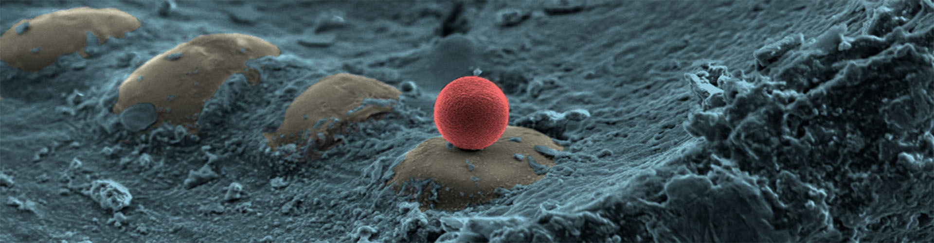
Adelaide Microscopy provides high to ultra-high-resolution SEM capabilities for both biological and materials science applications. The facility supports:

  • High-resolution surface imaging down to the nanometre scale
  • Automated mineralogy for geological and mining applications
  • Elemental analysis using energy-dispersive X-ray spectroscopy (EDS)
  • Crystallographic analysis via electron backscatter diffraction (EBSD)
  • Cathodoluminescence imaging for optical property studies
  • Electron beam induced current (EBIC) 
  • Focused ion beam (FIB) milling for site-specific cross-sectioning and 3D reconstruction as well as TEM sample preparation
  • Variable pressure for imaging non-conductive or hydrated samples
  • Structural imaging using STEM and selected area diffraction (SAD). 

These instruments support a wide range of applications, including nanotechnology, materials science geology, environmental, space and defence, and biology and biomedical research. 

Transmission Electron Microscopy

Adelaide Microscopy hosts several high-performance TEM instruments for atomic-scale imaging and analysis of biological and material samples:

  • Aberration Corrected TEM
  • Dedicated Cryo-TEM 
  • Materials TEM
  • Biological TEM.

These instruments support a wide range of applications across materials science (nanomaterials, semiconductors, mineralogy) and biology (ultrastructure, single particle analysis), with expert staff available for training and support. 

X-Ray Instrumentation

Adelaide Microscopy offers a wide range of X-ray-based instruments for imaging, elemental analysis, and surface characterisation.  Instruments include:

  • X-ray Fluorescence Microscopy (XFM) - Arriving early 2026
  • X-ray photoelectron spectroscopy (XPS) 
  • X-ray Micro-Tomography (Micro-CT)
  • X-ray Diffraction (XRD).

These instruments support applications in materials science, geology, biology, and medical imaging, with expert staff available for training and support.

Mass Spectrometry

Adelaide Microscopy offers advanced mass spectrometry instrumentation for detailed isotopic, elemental, surface and molecular analysis, including:

  • Laser Ablation ICP-MS
  • Solutions ICP-MS 
  • TOF-SIMS.

These facilities support research across geochemistry, biomedicine, environmental science, and materials science

Confocal

Adelaide Microscopy provides access to several advanced confocal microscopy systems designed for high-resolution imaging of biological and material samples. Their instruments support:

  • Super-resolution imaging 
  • Multiphoton microscopy for deep tissue imaging. 
  • Spectral detection for fluorescence analysis.
  • Live-cell imaging and dynamic studies.
  • Surface roughness and shape measurements. 

These capabilities are located across several nodes and are suitable for researchers across life sciences, materials science, and biomedical engineering, offering both routine imaging and cutting-edge techniques.

Optical Microscopy

Adelaide Microscopy offers a range of advanced optical microscopy systems for biological and materials science applications.

  • Lightsheet microscopy
  • Widefield Fluorescence  
  • Brightfield, Darkfield, Phase Contrast and Differential Interference Contrast (DIC)
  • Polarisation Microscopy.

These techniques are generally used in conjunction with advanced sample preparation and imaging workflows, supporting research in nanotechnology, biomedical science, and materials engineering and mineral identification. 

Preclinical Instrumentation

Adelaide Microscopy offers a suite of preclinical imaging instruments designed for in vivo studies in biomedical research. These instruments enable non-invasive, high-resolution imaging of live animals and biological samples, supporting research in areas such as drug development, disease progression, and anatomical studies. 

Sample Preparation

Adelaide Microscopy offers extensive sample preparation equipment across multiple facilities. These tools support both biological and materials science research including equipment designed for the following types of samples:

  • Histology 
  • Electron Microscopy (SEM and TEM) 
  • Live cell incubation
  • Mass Photometer
  • Ion Milling.

These instruments support preparation for SEM, TEM, optical microscopy, and advanced imaging techniques.

Data, Image Analysis and Visualisation

Adelaide Microscopy can assist with processing, quantification and visualisation of micro and nano-imaging data for a diverse range of applications - ranging from geological, chemical, biological, physical, materials, medical, dental and environmental, with clients from all scientific and engineering disciplines.

All major instrumentation at Adelaide Microscopy is charged at an hourly rate. The rate charged will depend on the instrument used and your status within Adelaide University.  We encourage users to operate the instruments themselves; however, an Adelaide Microscopy staff member can provide this service for an additional fee (referred to as an 'Operator' charge). Technical advice and assistance are provided free of charge. A sliding scale subsidy is offered to all government funded research and the charge categories are:

Adelaide University

Division of Research & Innovation subsidised internal rates apply to Adelaide University Staff, Students, and Affiliates (including Visitors, Adjunct, Clinical and Honorary Appointments), where project grant funding is administered by the Adelaide University and/or evidence of your Adelaide University Affiliate status is provided.  To qualify for this rate, an Adelaide University project code or evidence of your affiliate titleholder or visitor status must be provided.

Important notice regarding instrument charging

Please be advised that the project code provided at the time of booking will be used for instrument charging.  It is the researcher's responsibility to notify Adelaide Microscopy of any changes to this project code before the end of the month in which the instrument was booked.

Other institutions (or Government organisations) 

Associate rates are available to Australian publicly funded researchers and Government organisations.

Corporate & industry

Commercial entities should contact an Adelaide Microscopy staff member to discuss their microscopy needs. Adelaide University Staff or Students who are undertaking microscopy work on behalf of a commercial entity should discuss this with the Instrument Manager prior to commencing the work.

Please contact Adelaide Microscopy via email microscopy@adelaide.edu.au for our fee list.

Registration and booking

The equipment housed at Adelaide Microscopy is accessible to everyone (*conditions apply), but your booking options and charge rates may vary depending on your access level.

New enquiry?

To request access to our facility or enquire about the services we offer, please complete and submit the following form by clicking on the button below.  

Submit your analysis enquiry.

Existing users?

If you have registered and been trained on an instrument, log in to the Adelaide Microscopy Equipment Booking system using your Adelaide University ID, select University login via University login .

If you do not have an Adelaide University ID, log in via External user login .

A valid project is required by anyone wishing to book equipment or purchase consumables.

Registration information

Provide as much information as you can about your samples and aims of research so that we can attend to your request in a timely manner.

On receipt of your enquiry, we will put you in contact with one of our Microscopists/Microanalysts who will organise a face-to-face meeting to discuss your needs and ensure the right techniques are used to achieve maximum results.  Qualified Adelaide Microscopy staff are responsible for training all users for all instruments and laboratory procedures.

For most microscopy instrumentation, an initial training session will usually take around 1-4 hours in which your own sample is used. Prior to this session we will advise you on the most appropriate sample preparation technique.  It is important that you have your samples well prepared. This can be the most time consuming and critical aspect of your microscopy as it will affect the end result.  Instrument use is charged at the hourly rate depending on category.

All users are required to comply with University and local area Health, Safety and Wellbeing and must read the Adelaide Microscopy Health, Safety and Wellbeing Induction and Safe Operating Procedures (SOPs) prior to accessing the Centre and equipment.  Specific training courses are also offered by Adelaide Microscopy.  Any visitors accompanying you are required to report to sign in the Visitors book located at Adelaide Microscopy Reception on arrival.

Have a question?

Need help with booking?

Have a question or require assistance? Forgotten your password? Please email microscopy@adelaide.edu.au.

Contact us

Adelaide Microscopy

Location

Location
Adelaide Microscopy
Ground Floor NG13 
Helen Mayo North Building 
City Campus East, Adelaide SA 5005

Email

Tel: +61 8 8313 5855

Email

Email: microscopy@adelaide.edu.au